Impressive AO SLO images by MERLIN
Adaptive optics correction system has been integrated in the MERLIN device. It delivers AO-SLO images that enable to observe the retina with unprecedented level of details: the cellular level.
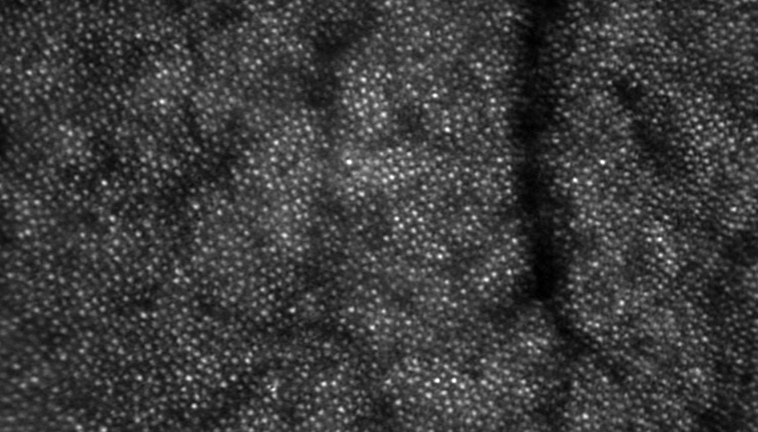
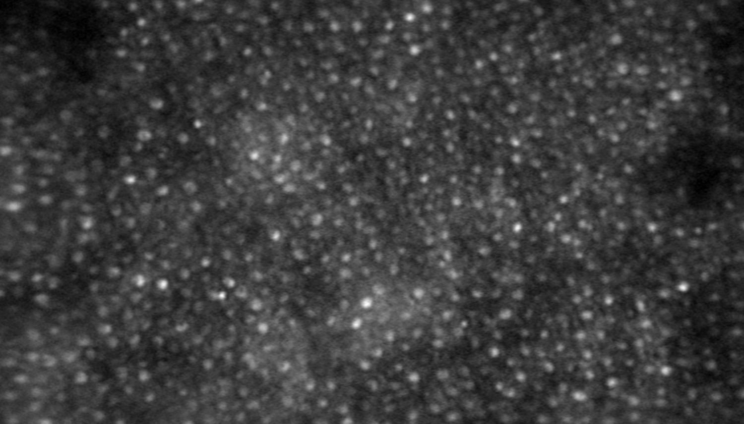
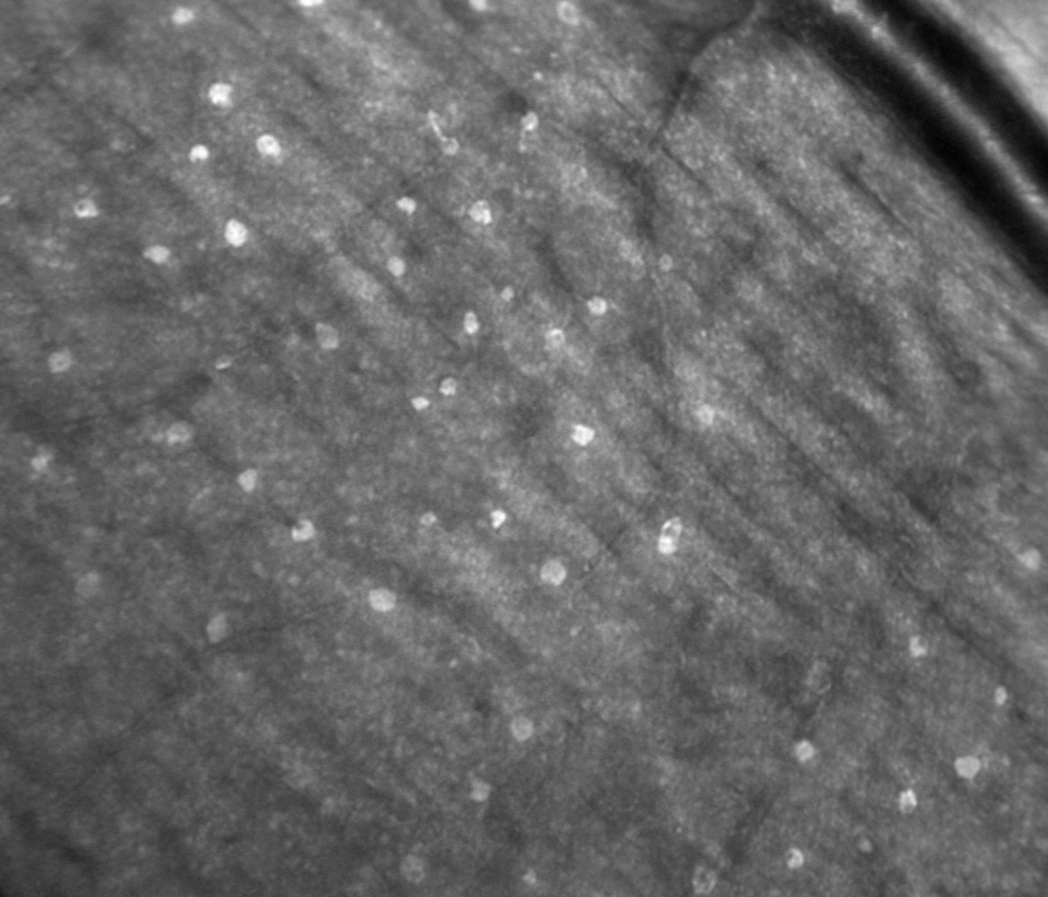
The following images were captured on healthy eyes. On each image, the structures observed are only a few micron in size.
- top image: individual visual cells (photoreceptor cones)
- middle image: more individual visual cells (photoreceptor cones and rods)
- bottom image: structure of blood vessels (artery in the corner, capillaries in the middle) as well as white cells (gunn dots).