Next-generation retinal imaging

The MERLIN project unites six academic and industrial partners who share a same ambition: improve the management of sight-threatening retinal diseases.

To do so, the MERLIN partners jointly develop a medical imaging technology that enables doctors to diagnose pathologies in the retina with unprecedented accuracy.

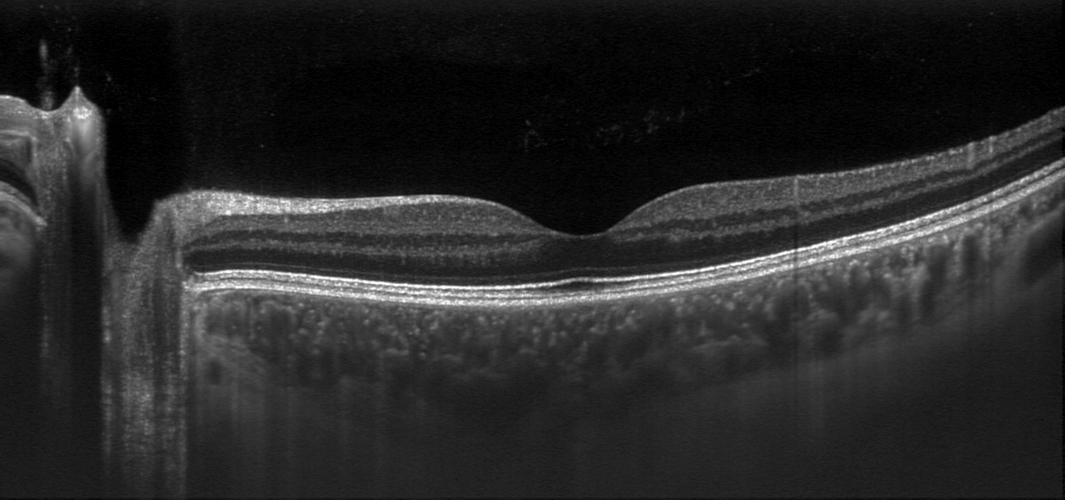
Tomographic overview image of the retina and choroid (OCT)

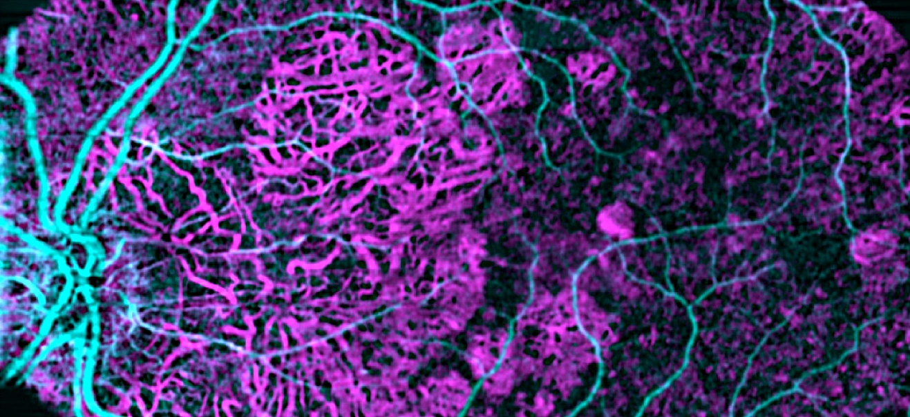
Overview image of retinal and choroidal microcirculation (OCTA) in a case of age-related macular degeneration

Enabling personalized medicine

The medical applications of MERLIN encompass a wide range of retinal diseases, including age-related macular degeneration (AMD) and inherited retinal dystrophies (IRDs), as well as chronic vascular conditions like diabetes. AMD and diabetic retinopathy (DR) are the leading causes of blindness worldwide in people over 55 years of age, while IRDs are the leading cause of vision loss in children.

As such pathologies progress slowly at the microscopic scale in the retina, both the clinical management of patients and the assessment of new treatments are currently difficult. Thanks to earlier detection and deeper phenotyping, MERLIN will enable the deployment of more timely, more personalized and more effective therapies.

Multimodal and multiscale imaging

In order to overcome current limitations in retinal imaging, the device developed in MERLIN will for the first time enable doctors to examine the back of the eye with multiple imaging modalities at both the macroscopic and microscopic scales.

The new instrumentation combines the most recent imaging techniques, including scanning laser ophthalmoscopy (SLO), optical coherence tomography (OCT) and OCT angiography (OCT-A), with new adaptive optics (AO) technology derived from astrophysics. As a result, the MERLIN device delivers stunning high-resolution images, which reveal previously invisible cellular and microvascular retinal details in 3 dimensions.

The project partners also develop image processing software for the analysis of microscopic pathological changes in the retina, and carry out clinical experimentations in order to evaluate and optimize both performance and usability in AMD and DR patients.
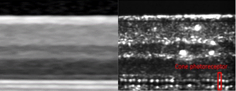
Close-up tomographic views of the retina : comparison between conventional OCT (left) and MERLIN AO-OCT (right) imaging technologies

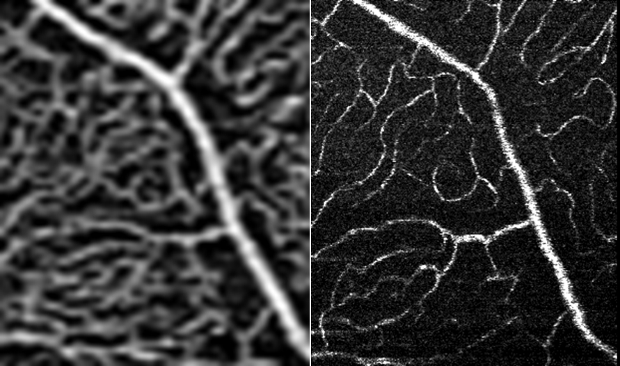
Close-up views of microcirculation in the upper retinal capillary plexus : comparison between conventional OCT angiography (left) and MERLIN AO-OCT angiography (right)

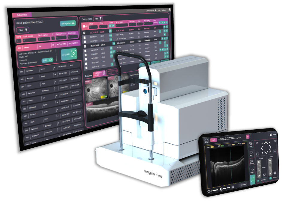
Design of the Merlin device hardware and software

Fast track towards clinical use

As the feasibility of MERLIN’s approach has already been demonstrated in a previous collaborative project (FP7 FAMOS, 2012-2017), the team’s effort is focused on transforming the technology from a preexisting laboratory prototype into a nearly commercial device usable in clinical trials.

After safety validation, performance and usability will be assessed in clinical settings at Paris Quinze-Vingts National Eye Hospital and Copenhagen Rigshopitalet using 2 clincial protoypes. Healthy volunteers and patients will be recruited under approved research protocols for examination with the Merlin device.

MERLIN partnersCountries
Imagine Eyes – project coordinatorFrance
Medical University of ViennaAustria
ICFO – The Institute of Photonic ScienceSpain
Erasmus Medical Center RotterdamNetherlands
INSERM & Quinze-Vingts National Eye HospitalFrance
Rigshospitalet & University of CopenhagenDenmark

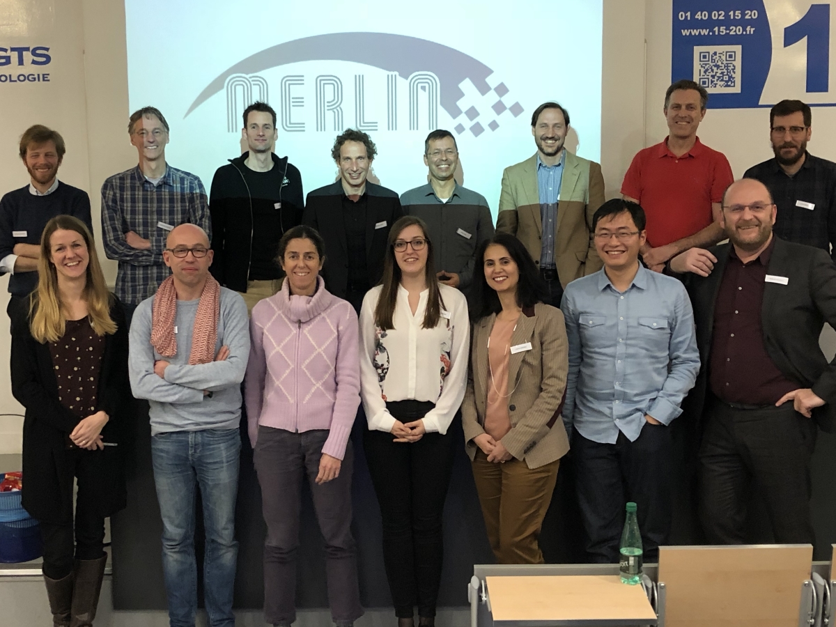
MERLIN partners at the project’s kick-off meeting in Paris

Supported by the European Commission

The MERLIN project has received € 4.9 M in funding from the European Union’s Horizon 2020 research and innovation programme under Grant Agreement No 780989.